
About Recurrent varicose veins
The circulatory system is made up of both veins and arteries. Arteries deliver fresh oxygen-rich blood from the heart to all tissues and veins then collect the oxygen-depleted blood again and transport it back to the heart.
The heart will pump blood through the arteries, but not all the way through the veins.
In the lower limbs, blood requires a different transport system to return it, against gravity, back to the heart. This transport system is made up of a pump again, the “calf pump” and one way valves within the veins.
It is for this reason that walking is a great way to keep your legs healthy, since the calf pump is activated all the time.
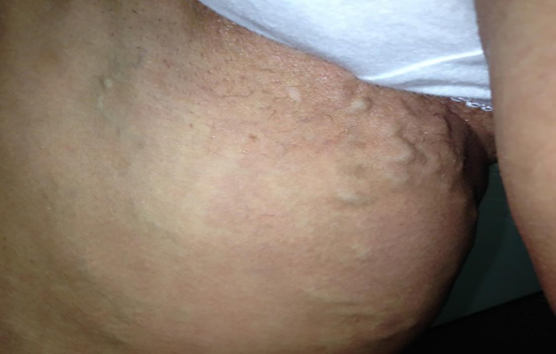
Broken one-way valves in the legs will cause blood to flow back through the vein and pool in the lower legs, this is known as venous reflux, and often presents as varicose veins. Varicose veins are large blue tortuous vessels that bulge from the surface of the skin.
Recurrent Varicose Veins
Why do varicose veins come back?
Varicose veins that return after previous treatment are common, and their reappearance does not necessarily mean that the original operation was performed badly. International specialists use the term REVAS (Recurrent Varices After Surgery) to describe this situation, and a consensus classification was developed in 1998 to distinguish between true recurrence, residual veins left behind at the first operation, and new veins caused by progression of the underlying disease.1
Understanding which of these is responsible in your particular case is the single most important step in planning successful treatment, and it can only be determined with a duplex ultrasound scan.1
The main causes of recurrence
Neovascularisation. This is the growth of small new blood vessels at the site where a vein was previously tied off, typically at the groin. A randomised trial found these new serpentine tributaries in 52% of legs after saphenofemoral ligation, making it the commonest single cause of recurrence in that study.2 It is thought to be a healing response to the trauma of surgical ligation, which is one of the reasons modern endovenous techniques that seal the vein from the inside, without a groin incision, have largely replaced stripping.
Residual veins. Veins that were refluxing at the time of the first operation but were not treated. This is more likely where surgery was planned on clinical examination alone rather than with a duplex scan, since the visible surface veins are only branches of the deeper refluxing trunks.
More than one source of reflux. Some patients have several separate points where reflux enters the superficial system. If only one was addressed, the others continue to feed varicose veins.
Progression of the disease. Venous disease is progressive. Valves that were competent at the time of the first treatment may fail later, producing genuinely new varicose veins in a leg that was successfully treated. This is not a failure of the earlier procedure.
Family history and individual risk. A strong family history of venous disease is common among patients presenting with recurrence, and other factors such as prolonged standing and higher body weight also contribute.
How common are recurrent varicose veins?
Reported recurrence rates vary very widely, from roughly 20% to 80%, depending on how recurrence is defined, how it is measured, and how long patients are followed.1 A recurrence detected on ultrasound is not the same as varicose veins a patient can see, and neither is the same as veins troublesome enough to need further treatment, so figures quoted from different studies are often not comparable.
In one randomised trial of stripping surgery, 62% of legs had developed clinically visible recurrent varicose veins by 11 years, although the great majority of those patients did not require another operation.3 In the same body of work, routine stripping of the great saphenous vein reduced two year recurrence from 43% to 25% compared with ligation alone.2
Recurrence rates after modern endovenous thermal ablation are generally lower than after open stripping surgery, and current international guidelines recommend endovenous ablation in preference to open surgery as first line treatment for superficial truncal reflux.4
How are recurrent varicose veins assessed?
Treatment of recurrent varicose veins should only be undertaken by a specialist in venous disease. These legs are anatomically more complex than untreated ones, and scar tissue from previous surgery changes the picture further.
Assessment begins with a thorough clinical examination followed by a detailed duplex ultrasound scan. Duplex ultrasound is the investigation of choice for recurrent varicose veins and is recommended by both European and American guidelines.14 The scan identifies where reflux is arising, whether the deep veins are involved, and which veins are feeding the visible varicosities.
Deep vein involvement matters. In a multicentre study of patients with recurrent veins after surgery, reflux in the deep veins was found in 27.4%.5 Patients with significant deep venous reflux, often following an earlier deep vein thrombosis, need a different treatment approach and sometimes are not candidates for treatment of the superficial veins at all.
From the scan an exact map is created, outlining every area that requires treatment. An individualised treatment plan is then drawn up and discussed with the patient before anything is done.
How are recurrent varicose veins treated?
Treatment is almost always a combination of techniques rather than a single procedure, tailored to the map produced by the duplex scan. Options include radiofrequency ablation of any refluxing trunk that remains, ultrasound guided foam sclerotherapy for veins arising from neovascularisation or deeper tributaries, and liquid sclerotherapy for the smaller surface veins.
Redo open surgery in the groin is generally avoided. Re-operating through scar tissue carries a higher risk of complications, and the evidence for repeat surgery in this setting is limited.1 Minimally invasive, ultrasound guided techniques are better suited to the altered anatomy and are performed in our office operating rooms under local anaesthetic.
Successful treatment of recurrent varicose veins demands patience and skill to achieve the best possible outcome, and treatment is often staged over more than one visit. Current guidelines advise reviewing patients at least three months after an initial ablation to assess whether further sclerotherapy or phlebectomy is needed.4
It is also worth being realistic about expectations. Because venous disease is progressive, treatment aims to control the current reflux and relieve symptoms rather than to guarantee that no vein will ever appear again. * Individual results may vary.
References
- Perrin MR, Guex JJ, Ruckley CV, et al. Recurrent varices after surgery (REVAS), a consensus document. Cardiovasc Surg. 2000;8(4):233–45. Journal
- Jones L, Braithwaite BD, Selwyn D, Cooke S, Earnshaw JJ. Neovascularisation is the principal cause of varicose vein recurrence: results of a randomised trial of stripping the long saphenous vein. Eur J Vasc Endovasc Surg. 1996;12(4):442–5. PubMed 8980434
- Winterborn RJ, Foy C, Earnshaw JJ. Causes of varicose vein recurrence: late results of a randomized controlled trial of stripping the long saphenous vein. J Vasc Surg. 2004;40(4):634–9. PubMed 15472588
- De Maeseneer MG, Kakkos SK, Aherne T, et al. European Society for Vascular Surgery (ESVS) 2022 clinical practice guidelines on the management of chronic venous disease of the lower limbs. Eur J Vasc Endovasc Surg. 2022;63(2):184–267. ESVS guideline
- Perrin MR, Labropoulos N, Leon LR Jr. Presentation of the patient with recurrent varices after surgery (REVAS). J Vasc Surg. 2006;43(2):327–34. PubMed 16476610
Further reading: What actually causes varicose veins?
Written and medically reviewed by Dr Johan Blignaut, MBChB (UOFS), FCS(SA)SURG, Specialist Surgeon.
Last medically reviewed: August 2026. This page is reviewed periodically and updated when clinical practice changes. It is general information, not medical advice – see our terms and medical disclaimer.